What Are The Big Causes Of Posterior Calcaneal Spur
Overview
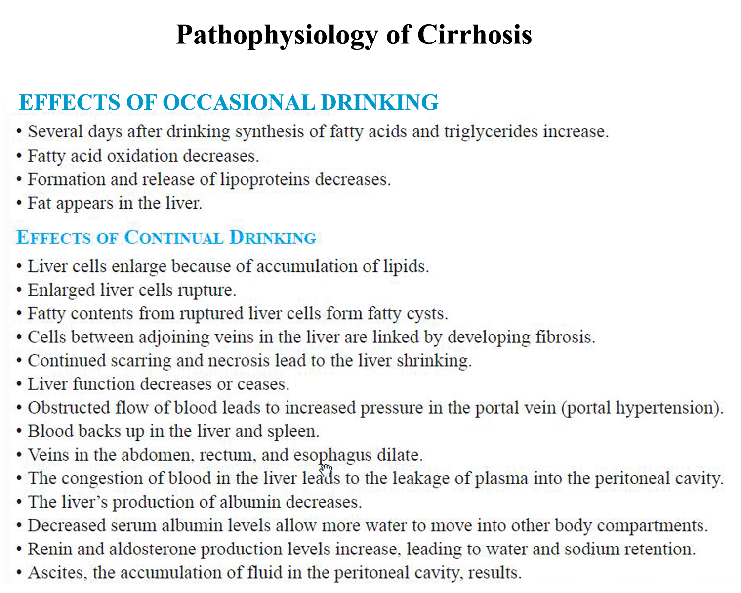
Heel spur is a hook of bone that forms on the heel bone. The condition itself is not the most painful; it is the inflammation and irritation that cause the heel pain. Often times, plantar fasciitis is a cause of heel spurs. When the ligaments are pulled away, calcium deposits form on the hooked bone. An orthotic will help relieve the pain associated with heel spurs.
Causes
The pain caused by heel spurs can be a sharp, stabbing pain when using the foot after a long period of rest. Sometimes it then reduces to a dull throb that can worsen when engaging in activities like jogging or jumping. People sometimes describe the pain of heel spurs and plantar fasciitis as a pin sticking into the bottom of the foot when they first stand up in the morning, this pain later turns into a bearable ache. The cause of the pain is generally not the heel spur itself, but the soft-tissue buildup associated with it. People often complain that the sharp pain returns after they stand up following sitting for a prolonged period of time.
Symptoms
The spur itself is not painful, however, if it is sharp and pointed it can poke into soft tissue surrounding the spur itself. As the bone spur irritates the tissue, inflammation and bruising can occur leading to heel pain. Heel spurs can affect your ability to do your usual work and/or activities, and can also trap and irritate the nerves in your heel area. They can change the way you walk, and can lead to knee, hip and low back injuries. If severe, they may require medical intervention.
Diagnosis
Most patients who are suffering with heel spurs can see them with an X-ray scan. They are normally hooked and extend into the heel. Some people who have heel spur may not even have noticeable symptoms, although could still be able to see a spur in an X-ray scan.
Non Surgical Treatment
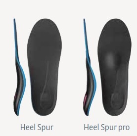
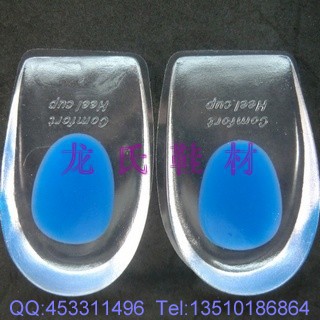
There are many temporary solutions to resolve the pain associated with irritation to the plantar ligaments. Common recommendations are ice and anti-inflammatory medications or even cortisone injections, however none of these solve the fundamental problem. To permanently resolve heel spurs you need to support and restrict the movement of the plantar ligaments. Flexible shoes will aggravate and often contribute to heel spurs. We recommend a RIGID orthotic that extends from the metatarsal heads to the heel to resolve heel spurs.
Surgical Treatment
Though conservative treatments for heel spurs work most of the time, there are some cases where we need to take your treatment to the next level. Luckily, with today?s technologies, you can still often avoid surgery. Some of the advanced technologies to treat a Heel Spur are Platelet Rich Plasma Therapy. Platelet Rich Plasma Therapy (also known as PRP) is one of several regenerative medicine techniques that University Foot and Ankle Institute has helped bring to foot and ankle care. This amazing in-office procedure allows the growth factors in the blood to be used to actually begin the healing process again long after your body has given up on healing the area. Heel Pain Shockwave Therapy. Shockwave therapy is a non-invasive procedure done in the office that allows for new blood to get to the region of fascia damage and help with healing. Results have been excellent with more than 70 percent of patients getting relief with only one treatment. Topaz for Heal Spurs and pain. Another minimally invasive technology technique is called Coblation Surgery using a Topaz probe. This minimally invasive procedure involves controlled heating of multiple tiny needles that are inserted through the skin and into the plantar fascia. This process, like PRP and Shockwave therapy, irritates the fascia enough to turn a chronic problem back into an acute problem, greatly increasing the chances of healing. Heel Spur Surgery. Endoscopic Plantar Fasciotomy is one surgical procedure that we consider to release the tight fascia. University Foot and Ankle Institute has perfected an endoscopic (camera guided) approach for fascia release to allow rapid healing and limited downtime with minimal pain.
Bursitis Of The Foot Pain Treatment
Overview
In the foot we have a unique situation in that between the shoes that we wear and the ground that we walk on various parts of the foot are constantly being ?micro? traumatized meaning that every time we take a step we do a small amount of damage to a particular part of the foot and eventually that part of the foot begins to hurt. The body?s response to this micro-trauma is to create a bursal sac to initially protect the area but if micro-traumatized enough the bursal sac itself becomes inflamed and we have a bursitis.
Causes
Inflammation of the calcaneal bursae is most commonly caused by repetitive overuse and cumulative trauma, as seen in runners wearing tight-fitting shoes. Such bursitis may also be associated with conditions such as gout, rheumatoid arthritis, and seronegative spondyloarthropathies. In some cases, subtendinous calcaneal bursitis is caused by bursal impingement between the Achilles tendon and an excessively prominent posterior superior aspect of a calcaneus that has been affected by Haglund deformity. With Haglund disease, impingement occurs during ankle dorsiflexion.
Symptoms
Symptoms of Achilles bursitis are often mistaken for Achilles tendinitis because of the location of the pain at the back of the heel. When you suffer from Achilles bursitis you will experience some or all of the following symptoms which are most noticeable when you begin activity after resting. High heels can add pressure on the retrocalcaneal bursa, subcutaneous calcaneal bursa, and Achilles tendon.
Diagnosis
When you are experiencing Achilles pain at the back of your heel, a visit to the doctor is always recommended. Getting a proper diagnosis is important so you can treat your condition correctly. A doctor visit is always recommended.
Non Surgical Treatment
Podiatric Care may include using anti-inflammatory oral medications or an injection of medication and local anesthetic to reduce the swelling in the bursa. An injection may be used for both diagnosis and for treatment. When you go to your doctor, x-rays are usually required to evaluate the structure of your foot and ankle to ensure no other problems exist in this area. They may advise you on different shoewear or prescribe a custom made orthotic to try and control the foot structure especially if you have excessive pronation. Sometimes patients are sent to Physical Therapy for treatment as well. To aid in relief of pressure points, some simple padding techniques can be utilized. Most all patients respond to these conservative measures once the area of irritation is removed.
Surgical Treatment
Surgery to remove the damaged bursa may be performed in extreme cases. If the bursitis is caused by an infection, then additional treatment is needed. Septic bursitis is caused by the presence of a pus-forming organism, usually staphylococcus aureus. This is confirmed by examining a sample of the fluid in the bursa and requires treatment with antibiotics taken by mouth, injected into a muscle or into a vein (intravenously). The bursa will also need to be drained by needle two or three times over the first week of treatment. When a patient has such a serious infection, there may be underlying causes. There could be undiscovered diabetes, or an inefficient immune system caused by human immunodeficiency virus infection (HIV).
Could Hammer Toe Lead To Soreness
 Overview
hammertoe can occur when feet are crammed into shoes so tight that the front of the toes are pushed against the front of the shoes for prolonged periods of time. One or more toes then remain bent with the middle knuckle pointing up, even when shoes are taken off. If the condition is left untreated and tight footwear is continually worn, these bent toes can become so rigid that they can no longer straighten out on their own. While any shoes that are too tight can lead to this condition, high heels seem to be a big culprit since the elevated ankle causes more weight to push the toes forward. This may explain why the condition affects more women than men.
Causes
Hammertoes are more commonly seen in women than men, due to the shoe styles women frequently wear: shoes with tight toe boxes and high heels. Genetics plays a role in some cases of hammertoes, as does trauma, infection, arthritis, and certain neurological and muscle disorders. But most cases of contracted toes are associated with various biomechanical abnormalities of the feet, such as flat feet and feet with abnormally high arches. These biomechanical abnormalities cause the muscles and tendons to be used excessively or improperly, which deforms the toes over time.
 Symptoms
Symptoms include sharp pain in the middle of the toe and difficulty straightening the toe. People with hammertoe may also develop blisters, which are fluid-filled pockets of skin, because the bent toe is likely to rub against the inside of a shoe. This increased friction may also lead to calluses, which are areas of thickened skin, and corns, which are hard lumps that may form on or between toes. Symptoms may be minor at first, but they can worsen over time.
Diagnosis
A hammertoe is usually diagnosed with a physical inspection of your toe. Imaging tests, such as X-rays, may be ordered if you have had a bone, muscle, or ligament injury in your toe.
Non Surgical Treatment
Treating hammertoe involves straightening the toe, making tendons in the toes flexible again, and preventing the problem from returning. Some simple treatments include splinting the toe to keep it straight and to stretch the tendons of the foot. Using over-the-counter hammertoe pads, cushions or straps to decrease discomfort Exercising the toes to relax the foot tendons (a session with a physical therapist may help you get started with foot exercises) Wearing shoes that fit properly and allow toes plenty of room to stretch out.
Surgical Treatment
For the surgical correction of a rigid hammertoe, the surgical procedure consists of removing the damaged skin where the corn is located. Then a small section of bone is removed at the level of the rigid joint. The sutures remain in place for approximately ten days. During this period of time it is important to keep the area dry. Most surgeons prefer to leave the bandage in place until the patient's follow-up visit, so there is no need for the patient to change the bandages at home. The patient is returned to a stiff-soled walking shoe in about two weeks. It is important to try and stay off the foot as much as possible during this time. Excessive swelling of the toe is the most common patient complaint. In severe cases of hammertoe deformity a pin may be required to hold the toe in place and the surgeon may elect to fuse the bones in the toe. This requires several weeks of recovery.
Treatment For Hammer Toes Pain
 Overview
What Is A Hammer toe? A hammer toe, or claw toe, describes a condition where the toe(s) become buckled, contracted or crooked. The toe could even cross over an adjacent toe, which is called a cross over toe. Any of the toes may be affected, but the 2nd and 5th toe are most commonly involved.
Causes
Medical problems, such as stroke or diabetes that affect the nerves, may also lead to hammertoe. For example, diabetes can result in poor circulation, especially in the feet. As a result, the person may not feel that their toes are bent into unnatural positions. The likelihood of developing hammertoe increases with age and may be affected by gender (more common in women) and toe length; for example, when the second toe is longer than the big toe, hammertoe is more likely to occur. Hammertoe may also be present at birth. Genetics may factor in to developing hammertoe, particularly if the foot is flat or has a high arch, resulting in instability.
 Symptoms
Pain on the bottom of your foot, especially under the ball of your foot, is one of the most common symptoms hammertoe associated with hammertoes. Other common signs and symptoms of hammertoes include pain at the top of your bent toe from footwear pressure. Corns on the top of your bent toe. Redness and swelling in your affected area. Decreased joint range of motion in your affected toe joints.
Diagnosis
Hammertoes are progressive, they don?t go away by themselves and usually they will get worse over time. However, not all cases are alike, some hammertoes progress more rapidly than others. Once your foot and ankle surgeon has evaluated your hammertoes, a treatment plan can be developed that is suited to your needs.
Non Surgical Treatment
Putting padding between your toes and strapping them in place can help to stop pain caused by the toes rubbing. Custom-made insoles for your shoes will help to take the pressure off any painful areas. Special shoes that are wider and deeper than normal can stop your toes rubbing. However if your pain persists your consultant may recommend an surgery.
Surgical Treatment
Several surgical procedures are available to the podiatric physician. For less severe deformities, the surgery will remove the bony prominence and restore normal alignment of the toe joint, thus relieving pain. Severe hammertoes, which are not fully reducible, may require more complex surgical procedures. Recuperation takes time, and some swelling and discomfort are common for several weeks following surgery. Any pain, however, is easily managed with medications prescribed by your podiatric physician.
Why Do I Get Hallux Valgus?
Overview
 A bunion is a bony growth in the lower joint of your big toe. It usually forms where the big toe pushes over against the second toe, forcing the joint to stick out. As a weight-bearing joint, this can be extremely painful. Calluses and blisters can form on the edge of the bunion, doubling the pain. This crippling foot affliction usually gets worse with time. Surgery for bunions can be complicated, expensive, painful and does not guarantee a well-formed foot as the outcome.
Causes
Bunions result from the long bone in the foot (metatarsal) and the big-toe bone becoming misaligned. The causes are likely to be a combination of genetics, wearing ill-fitting shoes, and the way that we walk or run. Arthritis sufferers are also prone to bunions.
Symptoms
Bunions are an often painful condition that may become even more painful as extra bone and a fluid-filled sac grow at the base of your big toe. Some of the most frequently experienced signs and symptoms associated with bunions, besides pain, include redness in your affected area. Blistering over your bunion. Callus formation around your bunion. Bursitis. Nerve damage (numbness and/or sharp pains) in your involved area. Bunions may also cause pain within and below your first metatarsophalangeal, or MTP, joint. Your bunion may become further dislocated and unstable as it progresses and may overload your adjacent joints.
Diagnosis
Your doctor is very likely to be able to diagnose your bunion simply by examining your foot. Even before that, he or she will probably ask about your family and personal medical history and evaluate the types of shoes you wear. You'll be asked about your symptoms, when they started and when they occur. You may also be asked to flex your toe so that your doctor can get an idea of your range of motion. He or she may order x-rays in order to determine the extent of your deformity.
Non Surgical Treatment
A hinged flexible bunion splint, can relieve pain by providing corrective arch support and releasing tension away from the inflamed joint. Change shoes! Avoid flip flops, high-heels and shoes with pointed, narrow toe-boxes. Medicine will not prevent or cure bunions. However, the use of over the counter anti- inflammatory medications can help. Bunion splints, pads and arch supports can help redistribute weight and move pressure away from the big toe. 
Surgical Treatment
If bunions are causing severe foot pain or inflammation and swelling that limits daily activities and doesn't improve with rest, medication and comfortable shoes, surgery may be required. More than 100 surgical options are available for painful bunions. Some realign the foot's anatomy by cutting notches from the metatarsal bone or the bone of the big toe. The bones can then grow back without the slant that promotes bunion growth. The operation is usually done on an outpatient basis, but afterward, you probably will have to stay off your feet for a few weeks. Recovery takes about six weeks. Surgery is not recommended for a bunion that doesn't cause pain.
Study Overpronation Of The Feet
Overview
Pronation describes a slight inward rolling motion the foot makes during a normal walking or running stride. The foot (and ankle) roles slightly inward to accommodate movement. Some people, however, over-pronate and roll more than normal. With over-pronation, the arch of the foot flattens and causes excessive stress and pressure on the soft tissues of the foot. Over-pronation is more common in those with flat feet, and can lead to foot aches and pain, such as plantar fasciitis, Shin Splints and Knee Pain.
Causes
A common cause of pronation is heredity - we can inherit this biomechanical defect. The second most common cause is due to the way our feet were positioned in the uterus while we were developing; this is called a congenital defect. In either instance, the following occurs in our feet during our development.
Symptoms
If ignored, overpronation can lead to complications such as hammer toes, corns and calluses, shin splints, hallux rigidus and many more foot and lower leg problems. Hammer toes appear when the toes are placed under too much pressure and the ligaments and muscles in the toes begin to reduce in size, leading to the curvature of the toes and making them look like little hammers. Overpronators can develop hammertoes if they don?t wear an appropriate pair of shoes. Corns and calluses also appear as a result of overpronation. They form in response to excess pressure, and overpronators may find that they have excessive hard skin on the balls of the feet and inside edge of the big toe. It is the body?s way of protecting against excessive forces and friction. They can be painful.
Diagnosis
Look at your soles of your footwear: Your sneaker/shoes will display heavy wear marks on the outside portion of the heel and the inside portion above the arch up to the top of the big toe on the sole. The "wet-foot" test is another assessment. Dip the bottom of your foot in water and step on to a piece of paper (brown paper bag works well). Look at the shape of your foot. If you have a lot of trouble creating an arch, you likely overpronate. An evaluation from a professional could verify your foot type.
Non Surgical Treatment
Treatment with orthotics will provide the required arch support to effectively reduce excessive pronation and restore the foot and its posture to the right biomechanical position. It should be ensured that footwear has sufficient support, for example, shoes should have a firm heel counter to provide adequate control.
Surgical Treatment
HyProCure implant. A stent is placed into a naturally occurring space between the ankle bone and the heel bone/midfoot bone. The stent realigns the surfaces of the bones, allowing normal joint function. Generally tolerated in both pediatric and adult patients, with or without adjunct soft tissue procedures. Reported removal rates, published in scientific journals vary from 1%-6%.
Is It Possible To Address Severs Disease In The Home?
Overview
Sever?s disease is the most common cause of heel pain in children aged 9 to 14. Sever?s disease results from stress placed on the growth plate of the heel bone. An excessive amount of running or other activities can cause inflammation around the growth plate, which results in pain. Rest, ice and orthotics and proper shoes are usually prescribed.
Causes
The cause of Sever's disease is not entirely clear. It is most likely due to overuse or repeated minor trauma that happens in a lot of sporting activities - the cartilage join between the two parts of the bone can not take all the shear stress of the activities. Some children seem to be just more prone to it for an unknown reason - combine this with sport, especially if its on a hard surface and the risk of getting it increases. It can be almost epidemic at the start of some sports seasons, especially winter. At the start of winter, the grounds are often harder, but soften later. Children who are heavier are also at greater risk for developing calcaneal apophysitis.
Symptoms
Pain symptoms usually begin after a child begins a new sport or sporting season, and can worsen with athletic activities that involve running and jumping. It is common for a child with Sever?s disease to walk with a limp. Increased activity can lead to heel cord tightness (Achilles Tendon), resulting in pressure on the apophysis of the calcaneus. This will cause irritation of the growth plate and sometimes swelling in the heel area thus producing pain. This usually occurs in the early stages of puberty.
Diagnosis
A physical exam of the heel will show tenderness over the back of the heel but not in the Achilles tendon or plantar fascia. There may be tightness in the calf muscle, which contributes to tension on the heel. The tendons in the heel get stretched more in patients with flat feet. There is greater impact force on the heels of athletes with a high-arched, rigid foot. The doctor may order an x-ray because x-rays can confirm how mature the growth center is and if there are other sources of heel pain, such as a stress fracture or bone cyst. However, x-rays are not necessary to diagnose Sever?s disease, and it is not possible to make the diagnosis based on the x-ray alone.
Non Surgical Treatment
Decreasing or stopping sport is necessary until the pain reduces. Let pain be your guide, as it decreases you can slowly return to all activities. To help settle inflammation use an ice pack or rub an ice cube over the
painful area for 5 minutes daily whilst pain persists. Wearing supportive trainers during the day can help to soften the impact of walking on the heel. Encourage a normal pattern of walking. Complete the stretches below every day and before and after activity until your symptoms settle.